Your breasts are constantly evolving throughout your life due to natural processes like puberty, weight fluctuations, and hormonal shifts. Understanding these natural, expected breast changes versus potentially concerning symptoms is vital for proactive breast health. While many variations in breast tissue are completely normal, remaining “breast aware” and prioritizing annual screening mammograms are the most crucial steps you can take for early detection and peace of mind.
Common Causes of Breast Changes
The underlying causes of most breast changes are often linked to fluctuations in hormones, which influence the breast’s composition of fibrous (connective), glandular (milk-producing), and fatty tissue.
Common causes of changes that are typically benign include:
- Menstruation: Hormonal shifts during the monthly cycle can cause breasts to swell or become tender before or during your period. Increased production of estrogen early in the menstrual cycle causes breast swelling and tenderness in many women as breast ducts become larger.
- Hormonal medication: Medications such as birth control or hormone replacement therapy can alter breast density. Taking combined hormone therapy to relieve menopausal symptoms can also raise breast cancer risk.
- Pregnancy: Elevated levels of estrogen and progesterone cause breasts to increase in size and become tender during pregnancy. The mammary glands and milk ducts prepare to produce and carry milk, which can make breasts feel lumpy and full. Nipple discharge and bumps on the areolas may also occur.
- Perimenopause: In the years leading up to menopause, fluctuating estrogen and progesterone levels can cause breasts to feel more tender or lumpy. Changes in breast size and shape may also occur during this time, typically around age 45.
- Dietary factors: Consuming a high-fat diet and taking in excessive caffeine may contribute to breast pain or swelling.
What Are the Normal Changes in the Breasts as You Age?
As women age and progress through different reproductive phases, the composition and feel of their breasts naturally shift.
- Age and breast density: The combination of fibrous and glandular tissue is called fibroglandular tissue, which determines breast density. Breast density can change year over year and can only be measured through mammography. You are more likely to have higher breast density if you are younger (under 50). Dense breast tissue can make it harder to detect early-stage cancers on a mammogram and can also increase risk for developing breast cancer.
- Post-menopause changes: As women age past menopause, their breast tissue becomes less dense and fattier. Due to the loss of glandular tissue after menopause, breasts may become smaller and more elastic, making them feel less firm. However, weight gain during menopause can cause breasts to grow larger.
- Increased cancer risk with aging: The two biggest risk factors for developing breast cancer are being a woman and aging. Most breast cancers occur in women ages 55 and older. Starting menopause later than average can increase the risk of breast cancer. Experts recommend annual screening mammograms starting at age 40 (for women of average risk) and continuing as women age because the risk for breast cancer increases with age and hormonal breast changes.
What Breast Changes Should I Worry About That Can Be a Sign of Breast Cancer?
While many breast changes are benign, it is important to become familiar with how your breasts normally look and feel so you can quickly identify anything out of the ordinary. Routine monthly breast self-exams and clinical exams during your annual wellness visit can help you become “breast aware.”
If you notice any unusual or concerning changes, it is important to consult with your provider right away for further evaluation.
Breast symptoms that may warrant immediate attention include:
- A new lump in the breast or armpit that feels new or different; this could be a hard, painless mass or be soft, round, and painful.
- Thickening of tissue in the breast or armpit area.
- Swelling or changes in the overall size or shape of the breast.
- Changes to the skin’s appearance, such as dimpling, puckering, irritation, or flaky/red skin (dimpling or irritation in the breast skin can also resemble an orange peel).
- Nipple changes, including pain, a newly inverted or retracted nipple, or pulling in at the nipple.
- Nipple discharge that is clear, bloody, milky, watery, or green-black, and is not breast milk.
It is important to remember that most women with early-stage breast cancer will never develop visible or palpable symptoms until later stages. This is why early detection through annual screening mammograms is critical. Benign conditions, such as breast calcifications, breast pain, fibrocystic breast disease (which causes cysts) or mastitis, can cause symptoms that might be mistaken for breast cancer.
The most significant change to understand about breast cancer is that it often has no symptoms until it is more advanced. The two most common types of breast cancer are invasive ductal carcinoma, which starts in the milk ducts, and invasive lobular carcinoma, which begins in the milk-producing lobules.
The goal of annual screening is to detect these changes at the earliest possible stage:
- Early detection: Annual 3D mammography is valuable because it can detect breast cancer up to three years before a lump can be felt during a self-exam or clinical exam.
- Survival rates: Women diagnosed in the earliest stages (Stage 0 or Stage 1) typically require less invasive treatments and have a significantly higher chance of survival, with nearly a 100% five-year survival rate.
How Are Breast Changes Diagnosed?
If you observe any concerning breast changes or if your annual screening mammogram detects an area of concern, your physician may order advanced imaging technologies to further evaluate the tissue.
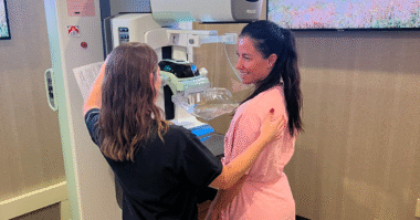
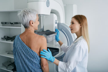
1. Annual Screening Mammograms
Annual 3D mammography (digital breast tomosynthesis) is the standard of care for women ages 40 and older at average risk for breast cancer. This technology creates multiple “slices” of breast tissue, forming a 3D reconstruction that allows radiologists to view breast tissue in layers. Annual 3D mammography has been clinically proven to detect breast cancer early and save 40% more lives. Annual screening is vital as it allows providers to compare images year over year, making it easier to detect subtle changes.
2. Diagnostic Mammograms and Follow-Up
If a change is detected during your annual screening, you may be “called back” for additional diagnostic imaging to further evaluate the concern. Only about 9% of women are recalled, and fewer than 1% will be found to have breast cancer, meaning the chances are much greater that the finding is negative. Alternatively, if you are experiencing symptoms like a worrisome lump or nipple discharge, your physician may refer you for a diagnostic mammogram.
A diagnostic mammogram appointment may include a combination of imaging technologies and procedures to obtain more detailed information about the area of concern.
3. Diagnostic Breast Imaging
Depending on the concern, the radiologist may use additional advanced imaging technologies:
- Breast ultrasound: This technology uses sound waves instead of X-rays to capture detailed images. It can be ordered to further evaluate specific areas of concern found on a 3D mammogram, for example, a lump that can be felt (palpable). Automated Breast Ultrasound (ABUS) is a supplemental imaging technology designed to help detect breast cancer in dense breast tissue, where it might be less visible on a mammogram. When ABUS is used with mammography, it has been shown to detect 3-4 additional cancers per 1,000 women screened.
- Breast MRI: Magnetic Resonance Imaging (MRI) uses a magnetic field and radio waves to produce images of soft tissue. If a change is detected through mammography, a breast MRI may be ordered for further evaluation or for additional surveillance between annual screening mammograms if a woman is considered high-risk. Breast MRI has been shown to detect 15-20 cancers per 1,000 women screened. Abbreviated Breast MRI (ABMRI) is a faster version of a breast MRI, typically used as supplemental screening for women with an average or lower risk for breast cancer or for those with dense breasts. ABMRI may also be used for women who qualify for, but are not able to proceed with, a longer breast MRI.
- Needle biopsy: If the radiologist identifies a concerning area through diagnostic imaging, a needle biopsy is a minimally invasive procedure performed to retrieve a small tissue sample for laboratory study to confirm if the tissue is benign or cancerous.
4. Breast Density Consideration
Breast density can complicate screening because dense tissue appears white on a mammogram, similar to cancerous areas, making abnormalities difficult to spot. Approximately 50% of the female population has dense breast tissue (categories C or D on the BI-RADS scale). Women with dense breasts have an increased relative risk of developing breast cancer. If you have dense breasts, supplemental screening with breast ultrasound, automated breast ultrasound, breast MRI or abbreviated breast MRI may be recommended in addition to annual 3D mammography.
Prioritizing Your Breast Health
Whether you are navigating normal age-related breast changes or evaluating a new or concerning symptom, maintaining your breast health over time is a priority. Charlotte Radiology offers comprehensive screening and diagnostic breast imaging services, staffed by board-certified, subspecialized radiologists and all-female technologists, to help support early detection and peace of mind.