Dense breast tissue can add a layer of complexity when it comes to evaluating and diagnosing breast cancer in women. The presence of dense tissue may make it more difficult to detect abnormalities in the breast and can be associated with an increased risk of developing breast cancer.
Dense breast tissue itself isn’t serious — in fact, it’s quite common. Almost 50% of women age 40 and older are found to have dense tissue on their mammogram results, according to the National Cancer Institute. You can’t get rid of dense breast tissue, but density can change year over year. Knowing more about breast density and the importance of annual 3D mammograms in identifying these changes can help you maintain your best breast health over time.
Read More: The Important Roles of 3D Mammography and Screening Breast Ultrasound in Early Detection
What Is Dense Breast Tissue?
Breasts are made of milk glands and ducts, connective tissue (also known as fibrous tissue), and fat, all of which make up your breast density. Breast density is essentially the ratio of glandular and connective tissue to fatty tissue.
Unlike lumps or changes in skin texture, you can’t feel or see dense breast tissue during a monthly breast self-exam. Your primary care physician won’t be able to determine your breast density by sight or touch, either. Only a mammogram can allow physicians to identify and categorize breast density.
Four Types of Breast Density
Breast density is measured using the Breast Imaging Report and Data System (BI-RADS) scale, which was created by the American College of Radiology. The levels of density are rated on a letter scale. The levels of the BI-RADS scale include:
- A: The breast is almost entirely fatty tissue. Around 10% of women have this level of breast density.
- B: The breast includes scattered areas of glandular and connective tissue, known as fibroglandular tissue, but most of the breast is fatty tissue. Around 40% of women have scattered density.
- C: Most of the breast contains dense glandular and connective tissue but there are some areas of fatty tissue, which is known as heterogeneously dense. About 40% of women have this level.
- D: The breast is almost entirely glandular and connective tissue, with fatty tissue comprising just a small portion. Known as extremely dense breast tissue, around 10% of women have this type.
Dense Breast Tissue Causes and Risk Factors
Researchers don’t know for sure what causes dense breast tissue, but family history seems to contribute. Many women with dense breasts inherit dense tissue from at least one of their parents.
Several factors can increase your risk for having high breast density, including:
- Being pregnant or breastfeeding
- Being younger (under 50)
- Having a lower body weight
- Taking hormone replacement therapy
The makeup of your breast tissue may not stay the same throughout your life. Breast density can change over time, and as you age or gain weight, your breasts may lose some of their density. For example, breast density often declines for women after menopause.
Read More: How to Prepare for an Annual Screening Mammogram
Dense Breasts and Cancer Risk
If you have heterogeneously dense or extremely dense breasts — categories C or D on the BI-RADS scale — you have a higher risk of breast cancer than women with mostly fatty breast tissue. Risk factors for breast cancer range from lifestyle choices, such as not exercising enough or drinking too much alcohol, to factors outside of your control, such as aging, genetic mutations and a family history of breast cancer. Researchers aren’t sure how dense breast tissue may contribute to the risk of breast cancer, but this tissue may contain more cells that can turn cancerous. Advanced 3D mammography is recommended to help better characterize changes in breast density and identify breast cancer early.
How Dense Breast Tissue Complicates Screening for Breast Cancer
Radiologists can have a tougher time detecting breast cancer in people with dense breasts because of how their breast tissue appears in their mammogram images.
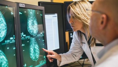
If you have mostly fatty breast tissue, tumors can’t really blend into the background. They stand out as white against the surrounding dark tissue, which makes them easier to detect. With dense breast tissue, however, potentially cancerous abnormalities in breast tissue aren’t as pronounced, especially early-stage. This is because even with advanced 3D mammography, areas of dense breast tissue, like tumors, appear white on a mammogram. As a result, cancers can “hide” and go undetected until they develop into later-stage breast cancer. Just because it’s an added layer of complexity doesn’t mean you should skip your regular mammogram, though.
The Importance of Annual Screening Mammograms
In women with dense breast tissue, 3D mammograms have been clinically proven to find most breast cancers, according to the American Cancer Society, and remain the standard of care for early detection. If you have dense breasts, keep up with your annual mammogram screenings starting at age 40 (or earlier, if you have a higher-than-average breast cancer risk) to detect subtle changes in your breast tissue or density over time.
3D mammographic imaging captures “slices” of the breast, layering the images to create a 3D view that is clearer and easier for a radiologist to read. Board-certified, subspecialized radiologists are highly trained in reading these scans to detect even the subtlest of changes. However, women with dense breasts may also benefit from supplemental screening options in addition to annual 3D mammograms.
In September 2024, the Food and Drug Administration began requiring patients’ mammogram reports to state whether their breasts were found to be dense or not dense and to explain the findings. Look for this information in your mammogram report and discuss any questions or concerns with your primary care physician as well as ask about adding supplemental screening options where recommended.
Advanced Imaging for Women With Dense Breast Tissue
If you have dense breasts, your physician may recommend other forms of breast imaging in addition to your annual 3D screening mammogram, including:
- Breast ultrasound. This form of imaging sends sound waves into the breast and produces images from their echoes. Breast ultrasound can help radiologists evaluate abnormal areas that may not show up clearly in a mammogram.
- Breast magnetic resonance imaging (MRI). Your physician may recommend an MRI of the breast, which forms pictures from radio waves and magnets, if a mammogram and ultrasound are inconclusive or they need to further characterize a change. In some cases, an MRI may show cancers that don’t appear in a mammogram. A breast MRI may also be added to a patient’s breast health plan, typically at six months following an annual mammogram for those at higher risk for developing breast cancer or with a previous breast cancer history.
- Abbreviated breast MRI (ABMRI): As the name implies, this is a shorter version of a breast MRI that focuses on the most vital images for early breast cancer detection. Charlotte Radiology offers ABMRI as supplemental screening primarily for women with dense breasts or other risk factors, in addition to annual 3D screening mammograms.
- Automated breast ultrasound (ABUS): ABUS is similar to screening breast ultrasound but uses a larger handheld paddle that the technologist hovers over each breast, taking multiple images. This supplemental screening exam is also available at Charlotte Radiology and is typically used for women with dense breasts or other risk factors, in addition to annual 3D screening mammograms.
These advanced imaging exams can improve breast cancer detection in women with dense breasts and have been shown in clinical studies to be effective when used in combination with mammography.


