Did you know that you could have a fractured spine without knowing it? A spinal compression fracture, also called vertebral compression fracture, can happen due to a traumatic event. However, many occur slowly due to bone loss that often comes with aging. These types of fractures may happen so slowly that you might only feel the effects many weeks later. Knowing the facts about spinal compression fractures can help you detect issues early before they affect your daily routine and quality of life.
Understanding a Spinal Compression Fracture
The spine consists of 33 vertebrae, which are small bones stacked on top of each other. Between each bone is an intervertebral disk that absorbs shock and helps to protect the bones. The spinal cord runs from the brain down through a canal in the vertebrae. Nerves branch off the spinal cord through openings along the sides of the vertebrae and travel outward.
Most of the weight in the body is supported by the front part of each vertebra, called the vertebral body. If a vertebral body becomes weak due to osteoporosis or cancer involvement, or if a large force is exerted on the bone during trauma, the bone can fracture and compress. Osteoporosis is a common condition, which results in bone weakness and is the most common cause of compression fractures. Many patients with osteoporosis present with a compression fracture after experiencing a fall. In some cases, osteoporosis is so severe that normal activities can cause a fracture to develop. Common situations include coughing, reaching up to a high shelf or twisting when stepping out of the shower.
Postmenopausal women are at the highest risk of developing a compression fracture. Approximately 25% of all postmenopausal women and 40% of women over age 80 have had at least one. However, older men also have an increased risk for compression fractures, especially if they have osteoporosis. Other factors that increase your risk of developing a compression fracture include:
- Alcohol use
- Being at a high risk for falls
- Calcium or vitamin D deficiency
- Decreased vision
- Early menopause
- Family history of compression fracture
- Frailty
- Having dementia
- Low body weight
- Not getting enough exercise
- Previous compression fracture
- Smoking or other tobacco use
Read More: What Is Your Diagnostic Imaging IQ?
Signs and Symptoms of a Compression Fracture
Compression fractures caused by trauma or that happen suddenly typically come with severe back pain. The pain is often described as sharp or stabbing and may last for weeks or months. The pain is commonly felt in the back but may be felt in front of the spine or on your sides. Most vertebral fractures affect the mid to lower back, but they can occur in the mid-chest.
In some cases, particularly without acute trauma, you may not have any symptoms right away. Patients may not know they have a fracture until it’s spotted on an X-ray or other imaging tests they have for unrelated reasons.
Apart from initial acute pain, vertebral compression fractures caused by osteoporosis can lead to chronic symptoms:
- Back pain that is worse with movement or standing and better with rest or lying down
- Loss of height, up to 6 inches over time
- Stooped over posture or humped back, also known as kyphosis, which can increase forces on the spine and lead to higher risk of subsequent compression fracture
If the compression fracture also causes pressure on the spinal cord or nerves, you may develop additional symptoms over time, including:
- Bladder or bowel control issues
- Leg weakness, difficulty walking
- Numbness, tingling or weakness, particularly in the legs
Get immediate medical attention if you experience back pain along with sudden:
- Fever
- Loss of bladder or bowel control
- Numbness or weakness
- Severe pain
Getting a Clear Picture of the Issues
If you’re experiencing possible symptoms of a spinal compression fracture, imaging technology can help get a clear picture of what’s happening in and around your spine. Several diagnostic imaging tests can help diagnose vertebral fractures and rule out other conditions. Your provider may order one or more of the following:
- X-ray — X-rays provide clear pictures of bones and other dense structures in the body. This scan is often the first test your provider will order to confirm a spinal compression fracture diagnosis.
- Computed tomography (CT or CAT) — CT scans provide a more detailed evaluation of the bones than X-ray. These scans can help your provider determine if you have a compression fracture and which portions of the bone are damaged
- Magnetic resonance imaging (MRI) — MRIs provide the best evaluation for compression fractures. They can show the location and anatomy of the fracture, as well as the amount of swelling that is present in the bone, termed marrow edema. MRI also offers the best view of the spinal cord and nerves to determine whether the bone is impinging on any of these structures.
- Dual-energy X-ray absorptiometry (DXA or DEXA) scan — A DEXA scan is a bone density test, that is the gold standard to evaluate for osteoporosis.
Compression Fracture Treatments: Kyphoplasty and Vertebroplasty
In many cases, people with spinal compression fractures heal within about three months with conservative, non-surgical treatments, including a back brace, rest or short-term use of pain medication. Anti-inflammatory drugs and muscle relaxers may also help with pain relief. However, if your pain persists, your provider may recommend other treatments.
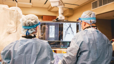
Two minimally invasive treatments for spinal compression fractures are kyphoplasty and vertebroplasty:
- Kyphoplasty — A needle is guided into the damaged vertebral body under imaging guidance. A balloon is then passed through the needle, which is inflated within the fractured bone to re-establish bone height and create space for controlled cement deposition. The balloon is then removed and a bone cement is instilled into the bone, which seals the fracture fractures together, improving pain, and preventing further height loss.
- Vertebroplasty — Cement is injected directly into the fractured vertebra without using a balloon to create additional space.
Our interventional experts at Charlotte Radiology will determine which treatment technique will produce the best outcome for each patient. People who receive kyphoplasty or vertebroplasty can typically return to their usual daily schedule right away with few restrictions.
Help You Can Trust
If you’re experiencing back pain or have signs of a possible spinal compression fracture, the experts at Vascular & Interventional Specialists can help. Our providers are trusted leaders in interventional radiology, offering leading-edge minimally invasive treatments for compression fractures. Talk to your primary care physician if you notice any symptoms and to see if an interventional radiology consultation is an option for you.


