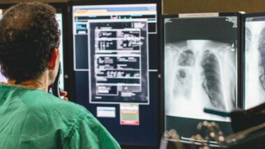
When a sports injury happens, it’s important to get a fast, accurate diagnosis, so you can get back in play as soon as possible. Doctors use imaging for many types of sports injuries to obtain a detailed picture of the injured area and develop the most effective treatment plan.
Depending on the type of injury you have, your doctor may use one of the following imaging techniques to help with diagnosis:
- X-rays can show skeletal system injuries, such as bone fractures and arthritis.
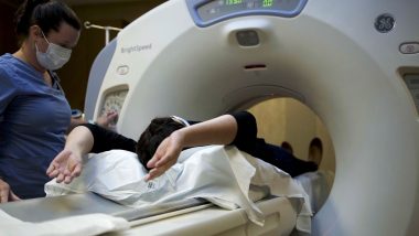
- CT scans create cross-sectional pictures of the body using X-rays. They are often used for traumatic injuries of the head, spine, chest and abdomen and CTs are also often used to better evaluate complex bone fractures.
- MRI scans use powerful magnets and radio waves to create images of soft tissues, such as the brain, nerves, muscles and organs.
- Ultrasound uses high-frequency sound waves to image muscles, tendons and internal organs.
A variety of common sports injuries often requiring imaging to help doctors establish a diagnosis.
Strains and Sprains
A strain happens when a muscle or tendon, a band of tissue that connects muscles to bones, stretches or tears. Repetitive motion and acute injury can lead to strains, making them common injuries for people who play tennis or golf. Sprains occur when ligaments, which connect the end of one bone to another, become stretched or torn, usually from a trauma such as a sudden blow or a fall. Ankles, knees and wrists are especially susceptible to strains and sprains. Hamstring strains, which affect the muscles at the back of the thigh, are also common for athletes who play sports that require sprinting.
To diagnose a sprain or strain and determine the severity of the injury, your doctor may start with an X-ray to rule out other causes of pain, such as broken bones. Your doctor may also order an MRI or ultrasound.
Read More: What Is Your Diagnostic Imaging IQ?
ACL and MCL Tears
Another type of ligament injury common in athletes is an anterior cruciate ligament (ACL) tear. People who play contact sports such as football, soccer and basketball are at higher risk for this type of injury, which happens when the ligament at the middle of your knee joint stretches and tears. Tears of the medial collateral ligament (MCL), which is located on the inside of your knee, are also common.
X-rays and MRI scans can help your doctor rule out other injuries and make an accurate diagnosis.
Meniscus Tears
A meniscus tear is another very common knee injury among athletes who play contact sports. Menisci are pieces of fibrocartilage that sit between the two bones that meet to form your knee joint. They provide cushioning and help stabilize your knee. When a meniscus tear occurs, you might feel a pop but still be able to play sports or exercise. However, the injury will worsen over the next few days. An MRI scan can help your doctor determine the extent of the damage.
Rotator Cuff Injuries
People who play sports that require an overhead arm motion, such as tennis, baseball and swimming, often sustain rotator cuff injuries. The rotator cuff is a group of muscles and tendons that hold your shoulder in its socket. Imaging such as MRI or ultrasound can help your doctor form a diagnosis.
Stress Fractures and Broken Bones
Broken bones often occur during sports, and they can result from a sudden injury, such as a fall or a blow, or from repeated stress over time, which is known as a stress fracture. A vast majority of bone fractures are best diagnosed and evaluated with X-rays. Some more subtle fractures from repetitive trauma which only extend part of the way across a bone, most commonly in the foot in runners, may not be appreciated on X-rays. When X-rays look normal and a doctor is concerned about a stress fracture that cannot be seen on an X-ray, a more sensitive MRI or nuclear medicine bone scan may be ordered.
Concussions
A concussion is a brain injury that can happen due to a sudden blow to the head, such as a hit in football or a header in soccer. If a concussion is severe, a doctor might use CT scans and MRI imaging to evaluate damage to the brain and check for problems including hemorrhages.
Tendinitis
Tendinitis is a condition in which tendons become inflamed, often from repetitive motion over time. Golfer’s elbow and tennis elbow are two types of tendinitis common to athletes. It can also occur in the shoulders, hips and knees. X-rays, MRI scans, and ultrasound imaging can help doctors diagnose this condition.
Shin Splints
Shin splints are more accurately described as “medial tibia stress syndrome” and usually develop in athletes who exert a lot of weight-bearing on the tibia, or bigger lower leg bone. Activities such as jumping, running, and weightlifting are common culprits. This entity is a stress injury or stress fracture of the tibia. If this injury is not seen on X-rays, and MRI or nuclear medicine bone scan may be ordered.