If you have or may have a health condition that affects your brain or head, your doctor may order a computed tomography (or CT) scan. This advanced imaging procedure helps assess or diagnose a wide range of conditions. A head CT scan is also used to guide many types of minimally invasive procedures to treat brain or head conditions.
CT scans are non-invasive procedures that allow your provider to get a close-up look at structures and areas of concern inside the body. These scans combine X-ray and computer technology to create 3D images of blood vessels, bones and soft tissues.
Your provider may order a head CT scan if you experience a head injury or if you have symptoms such as:
- Abnormal behavior
- Confusion
- Dizziness
- Fainting, also called syncope
- Headaches or migraines
- Unexplained changes in hearing or vision
CT Versus MRI for Head and Brain Conditions
Head CT scans are often a first choice for diagnostic imaging when you have a known or suspected head injury or condition affecting your brain. Other imaging technologies, such as magnetic resonance imaging (MRI) scans, can provide more detailed images to help better characterize a concern. However, CT scans have several benefits when it comes to conditions affecting the brain and head, including:
- CT scans are typically faster than MRI scans depending on the study.
- In most cases, if you move slightly during a CT scan, the scan does not need to be repeated.
- The 3D images created during a CT scan typically show an abnormal area’s location, shape and size more clearly than an MRI scan.
What to Expect During a Head CT Scan
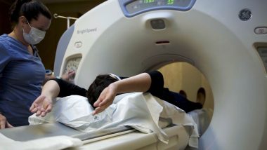
During a CT scan, you will lie on a table that slides into a donut-shaped opening in the CT machine. The radiology technologist will ask you to lay still and hold your breath for a few seconds while the machine works. The X-ray machine moves in a circular motion, taking 2D pictures of all sides of your body. The computer combines these detailed images to create a 3D view of the inside of your body.
You may have your CT scan done with or without contrast material, a special dye often given by injection or intravenously (IV). The dye causes blood vessels or certain tissue types to stand out in the images. After the contrast injection, you may feel flushed or warm or notice a slight metallic taste.
CT scans and contrast material are safe and generally do not have significant side effects. However, you will have a small amount of radiation exposure during a CT scan. Some people may have an allergic reaction to contrast dye. Rarely, contrast material may contribute to kidney damage. Before your scan, let your provider and radiology technologist know if you have any allergies, a history of kidney disease, or if you are pregnant.
Here are eight conditions that can be assessed or confirmed with a head CT scan.
Read More: What Is Your Diagnostic Imaging IQ?
1. Traumatic Brain Injuries
CT scans are commonly used to determine the severity of a traumatic brain injury (TBI) due to a blow or jolt to the head or body. Detailed images from CT scans can show brain bruising, bleeding and swelling following a head injury. They can also pinpoint the location of the damage to determine the best treatment options or to guide procedures used to treat TBIs.
2. Stroke
A CT scan, with or without contrast, is typically the first imaging procedure done within the initial hours or days after the symptoms of a stroke. CT scans can confirm a stroke diagnosis and rule out other conditions, such as abnormal tangles of blood vessels called arteriovenous malformations (AVMs), that may lead to future strokes. This information helps providers determine which treatment options will likely have the most significant effects.
CT scans can assess both ischemic strokes caused by blockages in blood vessels and hemorrhagic strokes caused by a brain bleed. They can also guide immediate medical interventions for a stroke.
3. Dementia
Dementia is a group of conditions that affect decision-making, learning, memory, problem-solving and thinking. The most common type of dementia is Alzheimer’s disease, but there are several other types. A CT scan can detect changes in the brain that are specific to different types of dementia. Knowing the type of dementia someone has can help them get the best treatment possible. CT scans can also rule out other conditions with symptoms similar to dementia or monitor the effectiveness of dementia treatments.
4. Tumors and Lesions
CT scans are commonly used to detect cancerous and non-cancerous brain tumors or lesions. The results of the scan may help:
- Confirm the diagnosis
- Develop treatment plans based on the characteristics, location or size of the lesion or tumor
- Monitor the effectiveness of treatment
- Plan treatment, such as radiation therapy
- Provide information about the characteristics and location of an abnormal growth
- Show changes in the lesion or tumor or a recurrence of cancer
- Stage cancer
5. Hemorrhage
In addition to strokes, a CT scan can find other types of hemorrhage, or bleeding, in the brain due to injury or another health condition. CT images can reveal the location and severity of the bleeding. The images may show one or more of the following:
- Epidural hematoma — bleeding between the skull and the outer covering of the brain, called the dura mater
- Intracerebral hemorrhage — bleeding within the brain tissue
- Subdural hematoma — blood pooling between the brain and the dura mater
CT images can help confirm the diagnosis of these conditions and develop a treatment plan.
6. Brain Aneurysm
A brain aneurysm, also called a cerebral aneurysm, happens when a weak spot in the wall of an artery causes it to bulge and fill with blood. Many aneurysms are small and do not cause serious issues. However, some aneurysms burst open, causing a life-threatening condition where blood spills into nearby tissue and prevents oxygenated blood from reaching other areas of the brain. Head CT scans with contrast injection or IV are often used to view blood flow in the brain and determine an aneurysm’s location, size and shape.
7. Skull Fractures
After head trauma, a CT scan can reveal skull fractures. The scan can help locate even small fractures that may lead to serious health concerns. Additionally, a CT scan can show bleeding, nerve or tissue damage or assess the severity of the condition following a known skull fracture.
8. Multiple Sclerosis
Multiple sclerosis (MS) is a condition that affects the brain, spinal cord and nervous system. In people with MS, the immune system attacks healthy material that helps protect nerve cells called myelin, affecting how effectively and quickly signals move between the brain and other parts of the body. Head CT scans may be used to detect lesions or other changes in the brain due to the progression of MS.
It’s important to talk to your physician immediately if you have an injury to the head and/or are experiencing worrisome symptoms impacting your ability to function. Your doctor can work with you to determine the next steps for diagnosis and treatment.