What are the differences between MRI and CT scans? Don’t feel bad if you struggle to distinguish between them — many people do. That’s because these key types of diagnostic imaging share a variety of similarities. Most CT scanners and MRI scanners are large tubes in which patients lie on a mobile table during exams. Both technologies are noninvasive and produce detailed images of many of the same parts of the body. Moreover, certain MRI and CT scans use contrast dye to make tissues or processes, such as blood flow, more visible.
Defining MRI and CT
Despite the many similarities between a CT and MRI exam, one key difference is how they produce images. MRI — also known as magnetic resonance imaging — uses a magnetic field and a radiofrequency current to create pictures based on the movement of protons in the body. It’s important to lie still during an MRI because movement can lead to blurry images.
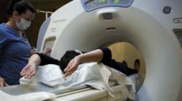
On the other hand, CT scans — another name for computed tomography —use a much different method to make capture images. A CT scan is a type of X-ray exam. While you lie still in the CT scanner, the machine rotates around you while aiming X-ray beams at your body. This allows the machine to produce image “slices” to form detailed 3D pictures.
Learn More: Estimate a Quote Today With Our Price Transparency Tool
What Can an MRI Scan Show?
MRI technology allows physicians to see bones, organs, and soft tissues, such as muscles, ligaments, and tendons. It can also show nerves and the spinal cord. Physicians use results from an MRI scan to:
- Diagnose sports injuries, such as sprained or strained muscles, torn anterior cruciate ligaments, and ruptured Achilles tendons
- Find benign and cancerous tumors
- Identify problems with the heart, lungs, spine, and circulatory and digestive systems
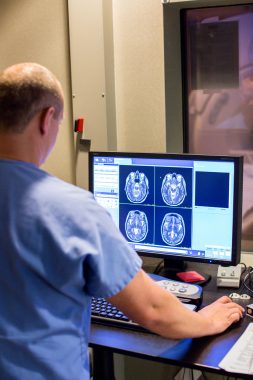
- Look for brain abnormalities, including tumors and aneurysms
- Pinpoint cartilage loss and bone diseases
- See the extent of joint inflammation
What Can a CT Scan Show?
Like MRI scans, CT scans can show many of the body’s internal structures, including bones, organs, and soft tissues. Physicians may use CT scans to see:
- Benign and cancerous tumors
- Blood clots
- Broken bones
- Emphysema
- Excess fluid in the lungs
- Injuries to organs
- Internal bleeding
- Joint damage
- Pneumonia
- Signs of heart disease
CT scans are especially valuable for diagnosing and understanding cancer, as well as determining cancer’s size and whether it has spread to other parts of the body, which is known as staging. Furthermore, CT scans help physicians determine whether a cancer treatment is working and see if cancer has returned.
Read More: CT Scans: What to Know
Key Differences Between MRI and CT Scan
There isn’t one scan that is better or more effective than the other. They each serve a purpose and depends on the area of the body or disease that needs to be examined. It’s important to understand the differences between an MRI and CT scan, including:
- Patient candidacy — You may not be able to have an MRI, which uses magnets, if you have a metal implant or an implanted device, such as a pacemaker.
- Speed — CT scans are faster than MRI scans, which is an advantage during emergencies. CT scans typically take 10 minutes or less, whereas MRI lasts, on average, 30–60 minutes, according to the American Academy of Family Physicians.
Whether you need an MRI or CT scan, you don’t have to go far. These types of imaging and more are available throughout Charlotte and the surrounding communities on an outpatient basis as part of Charlotte Radiology and Atrium Health’s joint venture, Carolinas Imaging Services. Our board-certified, subspecialized radiologists use their extensive training and expertise to review your images, giving you peace of mind and confidence in the results.
Learn more about outpatient imaging services.
Sources:
- CT general info — https://www.radiologyinfo.org/en/info/bodyct
- CT general info — https://medlineplus.gov/ctscans.html
- CT general info — https://www.nibib.nih.gov/science-education/science-topics/computed-tomography-ct
- MRI general info — https://medlineplus.gov/mriscans.html
- MRI general info — https://familydoctor.org/magnetic-resonance-imaging-mri/
- MRI general info — https://www.nibib.nih.gov/science-education/science-topics/magnetic-resonance-imaging-mri