When lower back pain becomes severe, it has the potential to affect your daily activities and quality of life. To diagnose the cause of your pain and find the right treatment, your doctor might order an MRI scan of your lower back, also known as a lumbar spine MRI. To help you prepare, discover what an MRI of the lumbar spine shows and what you can expect during the scan.
What Is a Lumbar Spine MRI?
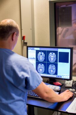
Magnetic resonance imaging (MRI) uses radio waves and a powerful magnetic field to create detailed images of the body’s soft tissues, organs, muscles and structures. A lumbar spine MRI focuses on the lower part of the spinal column, specifically:
- The five lumbar vertebrae from L1 to L5
- Coccyx (tailbone)
- Sacrum, the triangular bone connecting L5 to the coccyx
- Surrounding blood vessels, nerves, ligaments and tendons that support the spinal column
Read More: The Differences Between MRI and CT Scans
What Is a Lumbar Spine MRI Used For?
Your doctor might order a lumbar spine MRI if you have any of the following symptoms:
- Bladder or bowel incontinence
- Lower back pain that travels down the legs
- Lower back pain that doesn’t improve after several weeks of conservative treatment
- Weakness or numbness in the legs
Your doctor will use the results of imaging scans to examine the lumbar spine area and identify specific medical conditions, including:
- Compression or inflammation of the spinal cord and surrounding nerves
- Degeneration of joints, such as vertebral facet joints
- Disc herniation
- Infection of the discs, spinal cord and vertebrae
- Muscle strains or tears
- Spondylolisthesis, a condition in which a vertebra slips out of place
- Trauma to tissues
- Tumors
Additionally, MRI scans can be used to examine the spine after a surgical procedure or to evaluate people with certain conditions, such as:
- Brain or spinal cancer
- Multiple sclerosis
- Spinal birth defects
Read More: What Is Your Diagnostic Imaging IQ? Put Yourself to the Test
How to Prepare for an MRI Scan
After you confirm your appointment, you will receive instructions in advance on how to prepare for your MRI scan, including whether you need to fast beforehand. You will need to remove all metal accessories and digital devices, including jewelry, hearing aids, eyeglasses and electronic watches.
Your technologist will position you on a moveable exam table that slides in and out of the MRI machine. In some cases, especially if your doctor is looking for signs of infection or tumors, contrast dye is injected before the exam. This dye usually has no side effects, and allergic reactions are rare.
How Long Will a Lumbar MRI Scan Take?
Usually, a lumbar spine MRI takes about 30 to 60 minutes, including the time it takes to get in position. The exam is painless, but most MRI machines require you to be in an enclosed space. If you anticipate having anxiety, your doctor may be able to prescribe an anti-anxiety medication to take on the day of the exam. In this case, you will need to arrange for a ride home.
MRI Scans at an Outpatient Imaging Center
If your physician has ordered a lumbar spine MRI, you can decide where to have your imaging done. Choosing an outpatient imaging center like Carolinas Imaging Services offers many benefits, including:
- Advanced equipment
- Convenient hours that fit your schedule
- Multiple locations to choose from
- Next-day appointments
- Subspecialized radiologists
- Precise, reliable results
By getting fast, expert imaging you can trust, you will be on your way to getting the answers you need, allowing your doctor to diagnose your pain and get started on the right treatment plan for you.